Hyperdense Foci In Brain
How often have you read There are small scattered foci of signal abnormalities T2 hyperintensities or increased FLAIR signal in the cerebral white matter indicative of demyelinating disease chronic white matter ischemia due to microvascular disease or gliosis from an infectiousinflammatory disease process or words just like them in your MRI reports of your elderly. CT scan shows scattered hypodense foci in periventricular and subcortical white matter consistent with chronic small vessel ischemic disease.

A Ct Brain Showing A Well Defined Hyperdense Mass Lesion Located Download Scientific Diagram
You have basically two types of tissue in the brain.

Hyperdense foci in brain. Everyone is different and symptoms will vary in individual cases. What are the symptoms of brain lesions. Hyperdense MCA sign brain Pathology.
Multiple foci of T2FLAIR hyper intensity involving the subcortical white matterthere are a few punt ate periventricular white matter lesionsbilateral maxillary and ethmoid sinus mucosal thickening and there is layering fluid within the maxillary sinusesI need help understanding what this means and if any of it could be. It aids to improve psychological recall as well as performance to minimize forgetfulness. They can pose serious diagnostic problems which is reflected by their English name and abbreviation - UBOs Unidentified Bright Objects.
Hypodensityhyperdensity are features that usually are mentioned in MRI scans. T2-hyperintense foci are one of the most frequent findings in cerebral magnetic resonance imaging MRI. People often equate these bright spots with the potential diagnosis of multiple sclerosis MS or brain tumors but this is not necessarily the caseMedical professionals evaluate the spots based on a patients physical symptoms the location and size of the lesions and information gained from other tests.
Hyperdense Focus In Brain. Brain lesions can be caused by strokes as well as cancer. A lesion found in the blood is likely to have occurred because of a prior intra-arterial thrombosis.
From one-third to 80 percent of MRI scans performed on patients older than 65 show T2 hyperintense foci as of 2015. White matter hyperintensities WMHs are lesions in the brain that show up as areas of increased brightness when visualised by T2-weighted magnetic resonance imaging MRI. Foci on an MRI are periventricular white matter lesions evidence of changes in a patients brain that appear on the MRI as white spots states Timothy C.
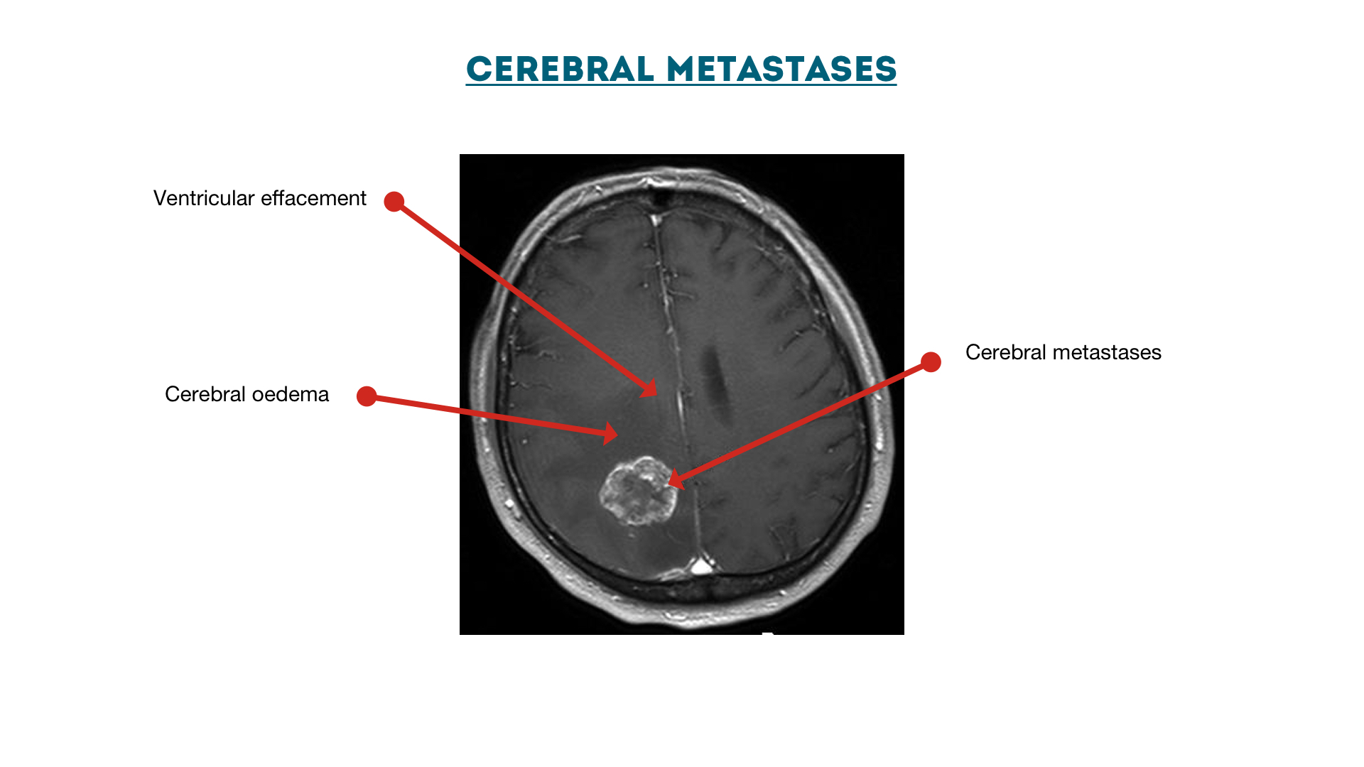
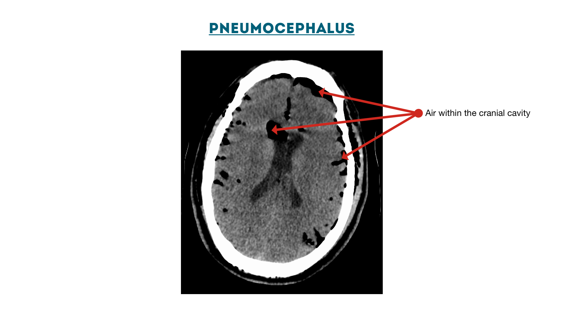
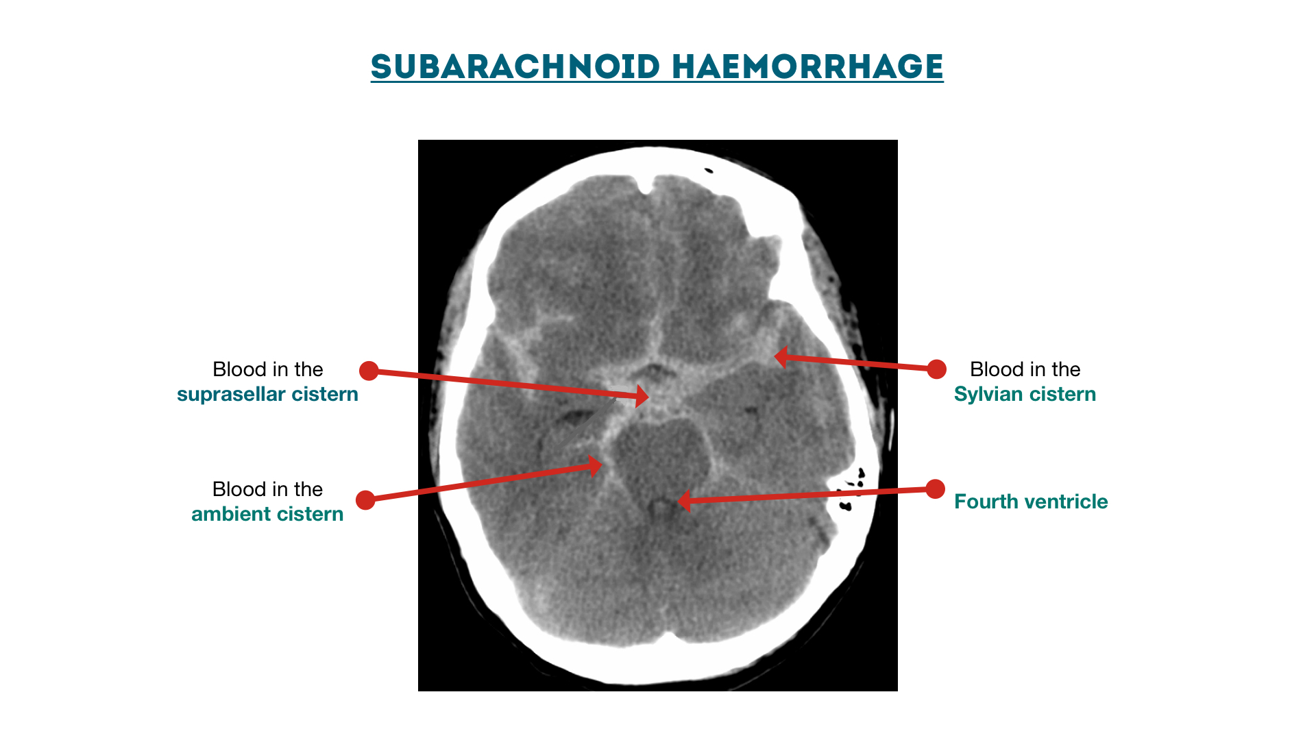
There is overlap between the entities with some cerebral metastases appearing in more than one list 1-6. This vital active ingredient is thought about water-soluble and also gives brain food which in turn boosts mental health and enhances metabolic process as well as acetylcholine natural chemical production. Hyperdense foci Hyperdensity on a CT head may be due to the presence of blood thrombus or calcification.
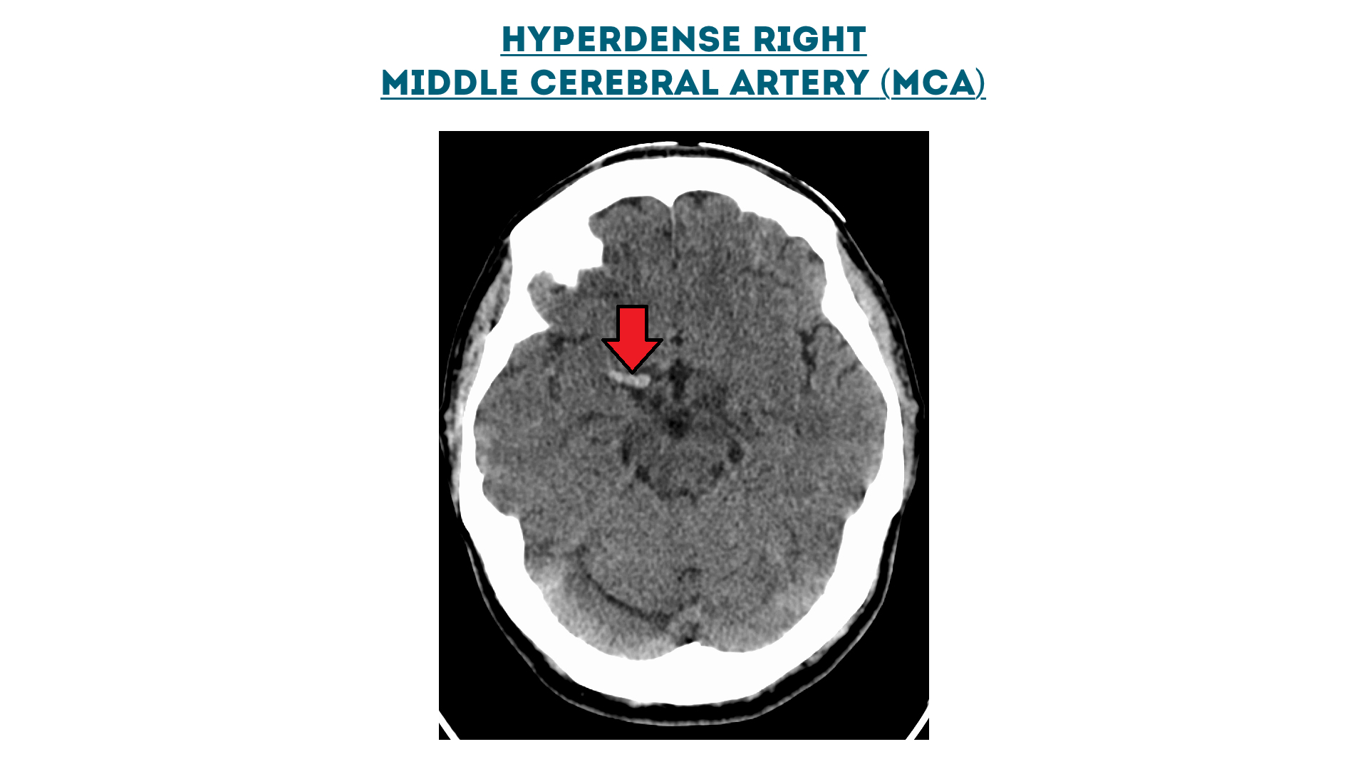
It is thus the earliest visible sign of MCA infarction as it is seen within 90 minutes after the event 1. Intratumoral hemorrhage also causes hyperdensity on CT images and is often associated with metastatic brain tumors glioblastomas pituitary adenomas and rarely with any of the other intracranial tumors. Symptoms of brain lesions vary depending on the type of lesion its extent and where it is found.
The gray matter rich in water and the white matter poor in water because rich in lipids such as myelin. What is white matter Hypodensity. The hyperdense lesion when found in other locations could be caused by personal injury viruses cancer and bacterial infections.
Hemorrhagic cerebral metastases mnemonic malignant melanoma can also be hyperdense when non-hemorrhagic due to melanin choriocarcinoma. The proximal portion of the MCA often extending into the terminal supraclinoid internal carotid. Also evidence of atherosclerosis in brain.
WMHs are also referred to as Leukoaraiosis and are often found in CT or MRIs of older patients. A hyperdense middle cerebral artery MCA is sometimes noted in total anterior circulation strokes TACS and indicates the presence of a large thrombus within the vessel. The hyperdense MCA sign refers to focal hyperdensity of the middle cerebral artery MCA on non-contrast brain CT and is the direct visualization of thromboembolic material within the lumen.
MRI allows to see water movement and water content in the brain using the proton magnetic spin. The hyperdensity of the arterial content is due to the thrombus having previously formed and contracted. The prevailing view is that these intensities are a marker of small-vessel vascular disease and in clinical practice are.
Tumors are also a cause of brain lesions and abnormal growth of brain cells. I received results from a brain MRI. Brain lesions are typically a symptom of this disease.
People with diabetes often have white matter foci.

Examples Of The Four Types Of Hyperdense Lesions On The Non Contrast Ct Download Scientific Diagram

Computed Tomography Imaging Revealed Hyperdense Foci Corresponding To Download Scientific Diagram

Brain Computed Tomography Showed A Hyperdense Nodule In The Right Download Scientific Diagram

A Ct Brain Showing A Well Defined Hyperdense Mass Lesion Located Download Scientific Diagram

Ct Head Interpretation Radiology Geeky Medics

Ct Head Interpretation Radiology Geeky Medics

Ct Scan Of The Brain Axial Sequence Shows An Area Of Hyperdensity In Download Scientific Diagram
Metallic Hyperdensity Sign On Noncontrast Ct Immediately After Mechanical Thrombectomy Predicts Parenchymal Hemorrhage In Patients With Acute Large Artery Occlusion American Journal Of Neuroradiology

Ct Scan Of The Brain Axial Sequence Shows An Area Of Hyperdensity In Download Scientific Diagram

Ct Scan Of The Brain Showing Bilateral Hyperdense Lesions In Basal Ganglia Download Scientific Diagram

Multiple Hyperdense Nodular Densities Due To Cerebral Granulomas In A Download Scientific Diagram

Brain Computed Tomography Showed A Hyperdense Nodule In The Right Download Scientific Diagram

Unenhanced Brain Computed Tomography A Shows Multiple Small Download Scientific Diagram

Case 2 Plain Ct Shows A Hyperdense Mass Lesion In The Left Frontal Lobe Download Scientific Diagram

Axial Non Enhanced Brain Ct Scan Revealed A Focal Small Hyperdensity In Download Scientific Diagram

Ct Head Interpretation Radiology Geeky Medics

Ct Scan Of The Brain Showing Bilateral Hyperdense Lesions In Basal Ganglia Download Scientific Diagram

A Ct Brain Plain Axial Image Showing Hyperdense Lesion In The Right Download Scientific Diagram

Ct Head Interpretation Radiology Geeky Medics
0 Response to "Hyperdense Foci In Brain"
Post a Comment